
- 1. Overview of Fractured Zygoma
- 2. The Close Connection Between the Zygoma and Oral Health
- 3. Zygomatic Implants – A "Lifesaver" for Dental Restoration
- 4. Combined Treatment Process: From Orthopedic Surgery to Dental Restoration
- 5. Leading Zygomatic Implant Brands
- 6. Post-Operative Care and Recovery
Modern medicine has revolutionized the way we approach facial trauma and subsequent dental rehabilitation. Fractures of the zygomatic bone often lead to significant aesthetic and functional challenges for patients. This article explores how advanced dental techniques provide hope for those suffering from severe facial bone loss.
1. Overview of Fractured Zygoma
Understanding the nature of zygomatic fractures is essential for choosing the right reconstructive path. These injuries can range from simple cracks to complex structural collapses.
1.1. Anatomy and Role in Facial Aesthetics
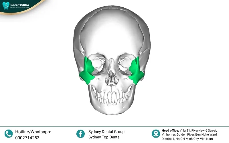
The zygomatic bone, commonly known as the cheekbone, serves as a primary pillar of the midface structure. It defines the facial contour and provides essential support for the surrounding soft tissues. A well-defined malar prominence is crucial for maintaining a youthful and symmetrical appearance.
Beyond aesthetics, this bone protects the delicate orbital contents and facilitates the attachment of powerful masticatory muscles. It forms the lateral wall and floor of the eye socket, ensuring ocular stability. Any displacement in this area can significantly alter a person’s facial proportions and vision.
The integration of the zygoma with the maxilla and temporal bone creates a robust framework for the entire skull. This connectivity allows the face to withstand the pressures of chewing and external impacts. Proper anatomical alignment is therefore vital for both functional health and psychological well-being.

1.2. Common Types of Fractures
Surgeons categorize zygomatic injuries based on the location and severity of the bone displacement. Various classification systems help medical professionals determine whether surgical intervention is necessary. These fractures often involve adjacent structures like the maxillary sinus or the orbital floor.
- Zygomatic Arch Fractures: Isolated arch fractures usually occur from direct lateral impacts to the side of the face. They can cause painful impingement on the coronoid process, making it difficult for patients to open their mouths.
- Zygomaticomaxillary Complex Fractures: This complex injury involves multiple fracture lines extending through the orbital rim and the maxillary buttress. It often results in a flattened cheek appearance and requires precise internal fixation for optimal recovery.
1.3. Recognition Signs
Identifying a zygomatic fracture early is critical for preventing long-term deformities and functional deficits. Patients often report specific sensory changes or physical alterations immediately following a facial impact. Clinical examination usually reveals a combination of visual cues and palpatory irregularities.
- Swelling and Deformity: Rapid edema and bruising around the eye can mask an underlying depression of the cheekbone. Once the swelling subsides, the loss of facial projection becomes more evident to the observer.
- Numbness and Restricted Movement: Damage to the infraorbital nerve often causes a persistent tingling or loss of sensation in the upper lip. Additionally, mechanical obstruction by the displaced bone can severely limit the patient’s ability to open their jaw.

2. The Close Connection Between the Zygoma and Oral Health
The health of the zygomatic bone is intrinsically linked to the stability of the upper dental arch. When this structure is compromised, the entire foundation for a healthy smile is put at risk.
2.1. How does zygomatic injury affect the bite?
A fractured zygoma can lead to a significant malocclusion where the upper and lower teeth no longer meet correctly. This misalignment occurs because the maxilla, which holds the teeth, is often displaced along with the cheekbone. Patients may experience difficulty chewing or feel that their jaw is “off-center.”
Persistent bite issues can cause chronic pain in the temporomandibular joint and lead to premature tooth wear. If left untreated, the uneven distribution of force weakens the supporting structures of the remaining teeth. Correcting the bony alignment is the first step in restoring a functional occlusal relationship.
Long-term functional impairment often requires a combination of orthognathic surgery and specialized dental prosthetics. Modern imaging allows clinicians to map the exact deviation and plan a precise corrective strategy. Reestablishing the proper bite is essential for the patient’s nutritional health and comfort.
2.2. Maxillary Bone Loss After Trauma or Long-term Tooth Loss
Severe facial trauma often triggers a biological process called resorption, where the jawbone begins to shrink. Without the stimulation of natural tooth roots, the bone loses its density and volume over time.
- Chronic tooth loss leads to a collapse of the alveolar ridge, making traditional dentures unstable.
- Extensive resorption in the posterior maxilla leaves very little bone available for standard dental implants.
This progressive bone loss creates a hollowed facial appearance and complicates any future restorative efforts. Understanding the rate of atrophy is crucial for timing the surgical intervention correctly.

2.3. Why do traditional tooth replacement methods fail when the zygoma is damaged?
Standard dental implants require a specific height and width of the alveolar bone to achieve primary stability. When the zygomatic-maxillary complex is damaged or atrophied, there is simply no foundation to hold the screw. Attempting to place implants in insufficient bone often leads to early failure and infection.
Traditional bone grafting procedures can be unpredictable and may require multiple surgeries over several months. Many patients find the long waiting periods and the risk of graft rejection to be major deterrents. Furthermore, extensive grafting might still not provide enough strength for a full-arch restoration.
Removable dentures often fail in these cases because they lack a stable surface to grip or rest upon. The lack of structural support from the zygoma means the denture can shift painfully during speech or eating. This loss of function significantly diminishes the patient’s quality of life and self-esteem.
3. Zygomatic Implants – A “Lifesaver” for Dental Restoration
Zygomatic implants offer a revolutionary alternative for patients who have been told they cannot have traditional implants. This specialized technique utilizes the dense bone of the cheek to provide a rock-solid foundation for teeth.
3.1. Concept of Zygomatic Implants
Unlike conventional implants that sit in the jawbone, zygomatic implants are much longer and extend into the malar bone. This bone is naturally dense and does not resorb like the alveolar bone in the mouth. By anchoring into this stable structure, the implants can support a full set of teeth.
The surgical technique involves a precise angulated placement to avoid the maxillary sinus and other vital structures. This approach bypasses the need for extensive bone grafting or sinus lifts in the majority of cases. It represents a significant leap forward in surgical prosthetic rehabilitation for complex patients.
Surgeons use advanced 3D planning software to visualize the optimal path for each implant before the procedure begins. This ensures maximum safety and allows for the immediate loading of a prosthetic bridge. The result is a highly stable and aesthetic solution that mimics natural dental function.

3.2. Indications
Candidates for this procedure usually have extreme bone atrophy that precludes standard dental implant placement. These individuals may have suffered from long-term denture wear or severe periodontal disease.
- Patients with previous facial trauma or failed bone grafts are ideal candidates for this approach.
- Individuals looking for a “Teeth-in-a-Day” solution despite having severe maxillary deficiency can benefit greatly.
Medical professionals evaluate the general health and bone density of each patient to ensure a high success rate. This procedure is specifically designed for those who have lost hope in traditional restorative methods.
3.3. Superior Advantages
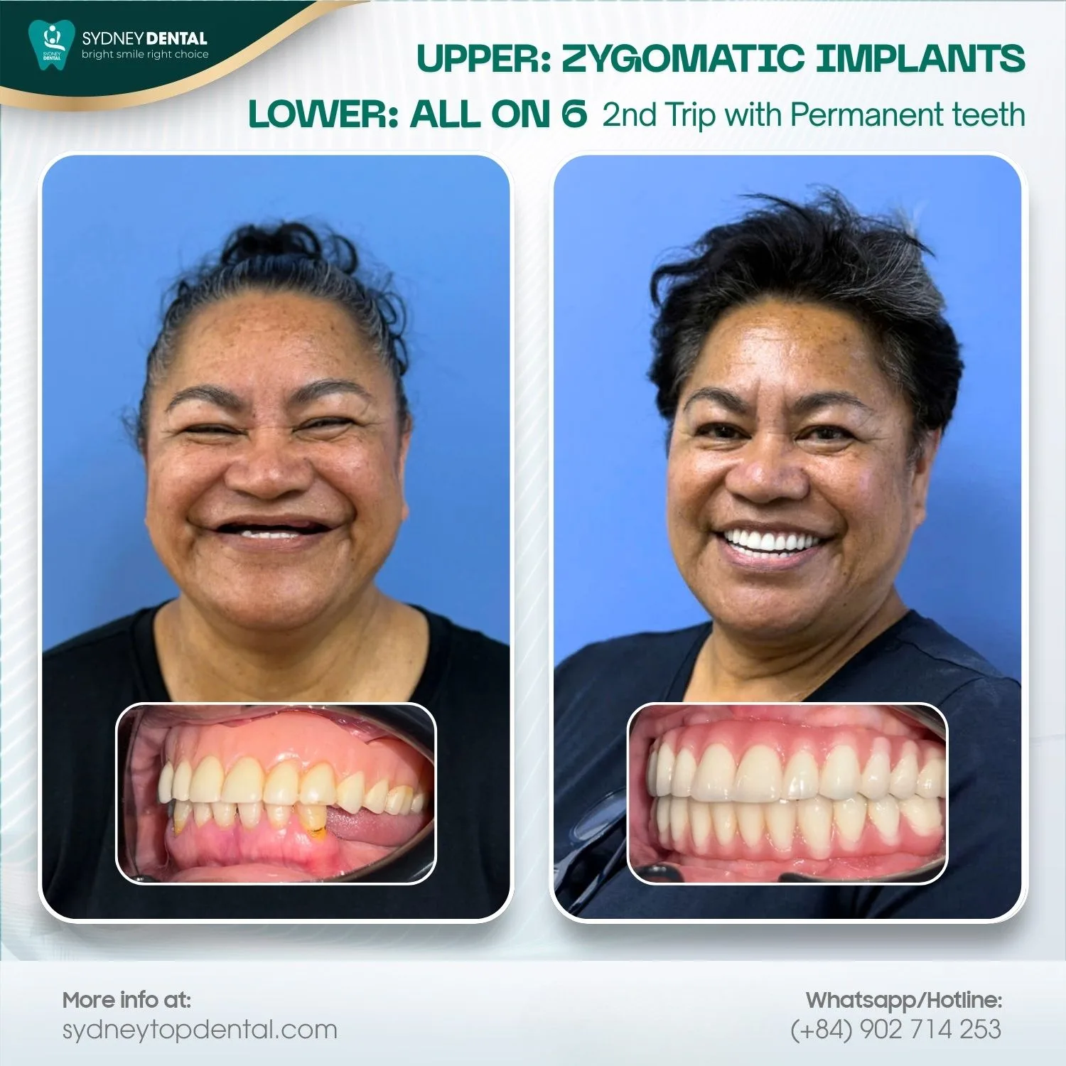
One of the most significant benefits is the elimination of the need for invasive bone grafting surgeries. This reduces the total treatment time and lessens the physical burden on the recovering patient.
- Patients can often receive a fixed temporary bridge on the same day as the implant surgery.
- The long-term success rates are exceptionally high due to the superior quality of the zygomatic bone.
This solution provides immediate functional and aesthetic improvements, allowing patients to eat and smile with confidence. It offers a permanent and durable fix for even the most challenging dental cases.
4. Combined Treatment Process: From Orthopedic Surgery to Dental Restoration
A successful outcome requires a coordinated effort between maxillofacial surgeons and restorative dentists. This multidisciplinary approach ensures that both the bone structure and the teeth are perfectly aligned.
Step 1: Examination and CT Cone Beam Scan
The initial phase involves a comprehensive clinical assessment to determine the extent of bone loss and structural damage. Specialized CT Cone Beam scans provide high-resolution 3D images of the midface and zygomatic regions. These images allow the surgical team to measure bone density and plan the exact implant trajectories.
Step 2: Facial Bone Reconstruction
In cases of recent or poorly healed fractures, the surgeon may first need to reposition the zygomatic bone. This procedure restores the anatomical framework and ensures a symmetrical base for the upcoming dental work. Small titanium plates or screws are often used to stabilize the bone segments during the healing process.
Step 3: Medical-Grade Zygomatic Implant Placement
The surgical team places the long zygomatic implants into the cheekbones using a specialized guided technique. This step is performed under general anesthesia or deep sedation to ensure the patient’s absolute comfort. The implants are carefully angled to provide the best possible support for the final dental prosthesis.
Step 4: Temporary Teeth and Final Restoration
Immediately following the surgery, a temporary bridge is usually attached to the implants to restore immediate function. Over the next three to six months, the implants integrate fully with the surrounding zygomatic bone. Once healing is complete, a final, custom-made prosthetic is fitted to provide a beautiful and lasting smile.

5. Leading Zygomatic Implant Brands
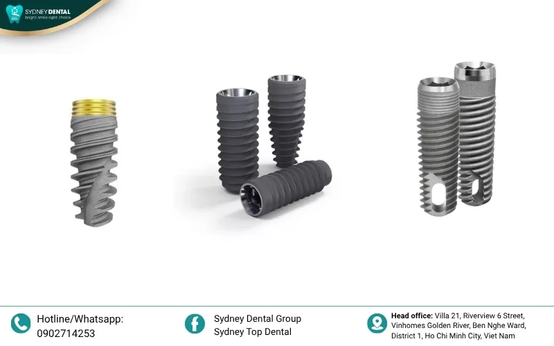
Choosing a reputable manufacturer is crucial for the long-term safety and reliability of the dental restoration. Leading brands invest heavily in clinical research and high-quality materials to ensure optimal osseointegration. These companies provide specialized components designed specifically for the unique geometry of the zygomatic bone.
- Nobel Biocare: A pioneer in the field, offering the most extensively documented zygomatic implant systems.
- Straumann: Known for their high-strength alloys and advanced surface treatments that promote faster bone healing.
- Zimmer Biomet: Provides versatile solutions for complex maxillofacial reconstructions and dental rehabilitations.
6. Post-Operative Care and Recovery
Proper aftercare is essential for ensuring the success of both the bone reconstruction and the implants. Patients must strictly follow a soft food diet during the initial weeks to avoid putting pressure on the surgical sites. Maintaining excellent oral hygiene is also vital to prevent infections during the critical early healing phase.
Surgeons typically prescribe a course of antibiotics and pain relievers to manage discomfort and reduce the risk of complications. Regular follow-up appointments allow the medical team to monitor the progress of osseointegration and soft tissue healing. Any unusual swelling or persistent pain should be reported to the clinic immediately for evaluation.
Long-term maintenance involves routine professional cleanings and check-ups at least twice a year. Using specialized cleaning tools can help keep the area around the implants free of plaque and debris. With diligent care, zygomatic implants can function effectively for many decades, providing a permanent health solution.
Restoring a smile after a zygomatic fracture requires a sophisticated blend of surgical skill and advanced dental technology. Zygomatic implants have transformed the lives of many by providing a stable foundation where traditional methods failed. By choosing the right specialists and brands, patients can achieve a functional and beautiful result that lasts a lifetime.
Sydney Top Dental
📍 Address: No. 21, Riverview 6 Street, Vinhomes Golden River Villas, Ben Nghe Ward, District 1, Ho Chi Minh City
📞 Hotline: 090 271 42 53
🌐 Facebook: Sydney Top Dental


Our Doctors
Get a Free Initial Consultation