
- 1. Why Do Zygomatic Implants Carry Higher Risks Than Standard Implants?
- 2. Common Biological Complications Based on Clinical Research
- 3. Late Complications: Risks Identified After 10-20 Years
- 4. How to Recognize Early Warning Signs of Failure
- 5. Expert Solutions for Preventing Complications
- 6. Zygomatic Implants: Minimizing Complications and Optimizing Outcomes by Dr. Hai
Zygomatic implants represent the ultimate solution for patients suffering from severe maxillary bone atrophy where traditional implants fail. However, undergoing such a complex procedure requires a deep understanding of potential biological and mechanical risks to ensure long-term success. Adhering to E-E-A-T principles, this guide provides a comprehensive overview of how to identify and prevent these clinical challenges effectively.
1. Why Do Zygomatic Implants Carry Higher Risks Than Standard Implants?
Zygomatic procedures involve navigating delicate facial structures that are not encountered during routine dental implant surgery. This inherent complexity significantly elevates the risk profile for patients with extreme bone loss.
1.1. Complex Anatomical Considerations
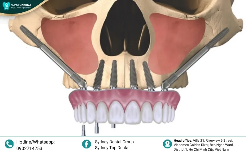
The zygomatic bone is located near vital structures including the maxillary sinus, the orbit, and various sensory nerves. Surgeons must precisely anchor long implants into the cheekbone while avoiding any damage to these sensitive areas. Any slight deviation in the drilling path can lead to immediate complications or long-term structural issues.
Furthermore, the path of the implant often traverses or bypasses the sinus cavity, which complicates the biological environment. Managing the interface between the oral cavity and the sinus requires meticulous planning and execution. Variations in individual facial anatomy make every case unique and inherently more challenging than standard alveolar placements.
1.2. Requirement for Exceptional Surgical Expertise
Performing zygomatic surgery demands advanced training in maxillofacial anatomy and specialized surgical techniques that go beyond general dentistry. A high learning curve exists for clinicians to master the blind-drilling aspects and proper prosthetic alignment required for success. Inexperienced practitioners may struggle with the spatial orientation necessary to avoid catastrophic failures in the midface region.
Precision is paramount because the long lever arm of a zygomatic implant magnifies any minor surgical or prosthetic error. Suboptimal placement can lead to excessive mechanical stress on the zygomatic bone or poor emergence profiles for the final teeth. Therefore, the outcome of the procedure is heavily dependent on the surgeon’s specialized skills and extensive clinical experience.
2. Common Biological Complications Based on Clinical Research
Clinical data from NCBI and long-term systematic reviews highlight several biological challenges that patients may face after surgery. Understanding these risks is the first step toward effective clinical management and patient safety.
2.1. Maxillary Sinusitis
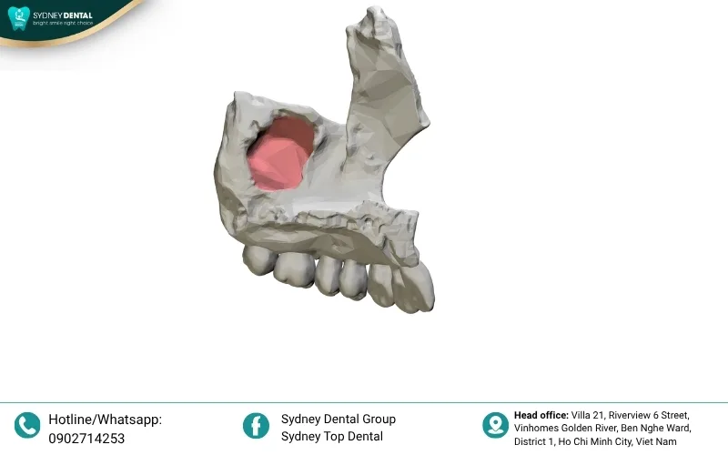
Maxillary sinusitis is identified as the most frequent complication, often occurring when the implant disrupts the sinus membrane or integrity. This condition can develop shortly after surgery or manifest years later as a chronic inflammatory response. Maintaining a healthy sinus environment is critical for the stability of the implant and the patient’s overall comfort.
Clinicians often utilize specialized techniques to protect the Schneiderian membrane and reduce the risk of secondary infections. Proper preoperative screening of the sinus health can significantly lower the incidence of post-surgical inflammatory issues. Monitoring patients for any signs of congestion or pressure is essential during the healing phase.
- Causes: Perforation of the sinus membrane during drilling; Infection spreading from the oral cavity; Pre-existing untreated chronic sinusitis.
- Symptoms: Persistent facial pressure or pain; Nasal discharge with an unpleasant odor; Recurrent localized headaches.
- Treatment: Targeted antibiotics and nasal decongestants; Endoscopic sinus surgery for refractory cases; Potential implant removal if infection persists.
2.2. Paresthesia and Nerve Damage
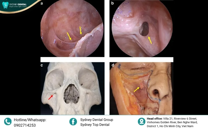
The proximity of the zygomatic implant path to the infraorbital nerve poses a significant risk for sensory disturbances. Damage can occur during the retraction of soft tissues or the actual placement of the hardware into the bone. While some sensory loss is temporary, permanent damage can severely impact the patient’s quality of life.
Surgeons must have a deep understanding of facial nerve distribution to navigate safely through the midface anatomy. Utilizing advanced imaging helps in identifying the exact location of nerve bundles before making any incisions. Continuous monitoring during the surgical procedure is a vital safety measure for every patient.
- Causes: Direct trauma to the infraorbital nerve; Excessive pressure from postoperative swelling; Incorrect implant positioning interfering with nerve pathways.
- Symptoms: Tingling or “pins and needles” sensation; Numbness persisting beyond the healing period; Altered sensitivity in the cheek or lip.
- Treatment: Conservative management with neurotropic vitamins; Surgical decompression if physical impingement is identified; Long-term neurological monitoring.

2.3. Peri-implantitis and Osseointegration Failure
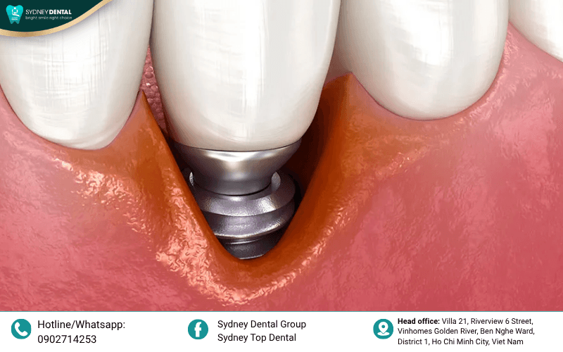
Peri-implantitis involves the inflammation of soft tissues and progressive loss of bone surrounding the implant. In zygomatic cases, this often starts at the crestal area where the implant emerges into the mouth. Without early intervention, the infection can compromise the entire stability of the zygomatic anchor.
Failure of osseointegration occurs when the bone does not successfully fuse with the titanium surface. This can result from overheating the bone during drilling or excessive loading before the bone has healed. A mobile zygomatic implant is a serious complication that usually necessitates total removal.
- Causes: Poor oral hygiene leading to bacterial accumulation; Mechanical overload of the bridge; Inadequate initial bone-to-implant contact.
- Symptoms: Bleeding or swelling of the gums; Visible bone loss on X-rays; Feeling of looseness in the dental bridge.
- Treatment: Deep mechanical cleaning and antimicrobial therapy; Bone grafting to repair localized defects; Explantation if stability is lost.

2.4. Oroantral Fistula
An oroantral fistula is an abnormal communication that develops between the oral cavity and the maxillary sinus. This complication often arises when an implant fails or if the surgical site does not heal properly. It allows bacteria from the mouth to enter the sinus, leading to chronic infections.
Closing a fistula requires a secondary surgical procedure to ensure a hermetic seal between the two cavities. Early detection is key to preventing the spread of infection into the deeper facial structures. Patients must be warned about the risks of smoking or blowing their nose forcefully during recovery.
- Causes: Failure of soft tissue closure; Infection leading to tissue necrosis; Removal of a failed implant without proper socket closure.
- Symptoms: Air or fluids passing from the mouth to the nose; Persistent foul-smelling discharge; Difficulty creating suction in the mouth.
- Treatment: Surgical closure using local soft tissue flaps; Extended courses of antibiotics; Strict adherence to sinus precautions.
3. Late Complications: Risks Identified After 10-20 Years
Systematic reviews covering two decades of clinical use reveal that complications can still emerge long after the initial procedure. These late-onset issues are often related to the wear and tear of the prosthetic components.
3.1. Mechanical Degradation of Prosthetics
The long-term mechanical stress on zygomatic restorations can lead to the fracture of screws or the framework. Because these implants support full-arch bridges, the biting forces are concentrated on only a few anchor points. Over time, the materials may fatigue and fail, requiring significant repairs or total replacement.
Regular maintenance and prosthetic adjustments are necessary to distribute forces evenly across the zygomatic anchors. Patients who clench or grind their teeth are at a significantly higher risk for these mechanical failures. Using high-strength materials and modern digital design can help mitigate some of these long-term risks.
- Causes: Material fatigue from functional loading; Improper bite alignment in the final bridge.
- Symptoms: Loosening of prosthetic screws; Visible cracks or fractures in the dental restoration.

3.2. Crestal Bone Loss Around the Implant Apex
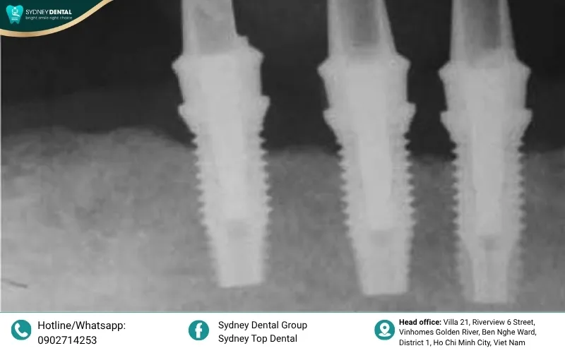
Although primary stability comes from the zygomatic bone, the health of the crestal bone is vital for soft tissue support. Progressive bone loss at the emergence site can expose the implant threads to the oral environment. This exposure increases the likelihood of infection and compromises the aesthetic result of the treatment.
Biological aging and changes in the jawbone structure can contribute to this slow recession over the decades. Clinicians must monitor the bone levels through annual X-rays to catch early signs of instability. Maintaining thick, healthy gum tissue around the implant neck is a key preventive strategy.
- Causes: Chronic low-grade inflammation at the gum interface; Natural physiological bone remodeling.
- Symptoms: Gradual recession of the gums; Increased food entrapment around the implant neck.
4. How to Recognize Early Warning Signs of Failure
Identifying potential issues in their early stages can often prevent a minor complication from becoming a total surgical failure. Patients must remain vigilant and report any unusual sensations to their clinical team immediately.
- Pain and Swelling: Persistent pain that does not subside with prescribed medication may indicate an underlying infection or improper implant seating. While some swelling is normal, any sudden increase in redness or heat in the cheek area is a significant concern. Patients should monitor the external facial tissues as closely as the internal surgical site.
- Pus and Odor: The presence of a foul odor or a salty discharge often points toward a sinus infection or a developing fistula. Pus formation around the gum line is a clear indicator of bacterial activity that requires immediate professional intervention. These symptoms should never be ignored, as they can lead to rapid bone loss.
- Persistent Numbness: While temporary numbness is common after major facial surgery, any loss of sensation that lasts longer than a few weeks needs investigation. This could suggest that the implant is putting pressure on a nerve or that surgical trauma occurred. Early neurological assessment can help determine if corrective action is necessary to prevent permanent damage.
Prompt communication with your surgeon is the most effective way to manage these early symptoms and protect your investment. Addressing minor signs today can save you from complex revision surgeries in the future.
5. Expert Solutions for Preventing Complications
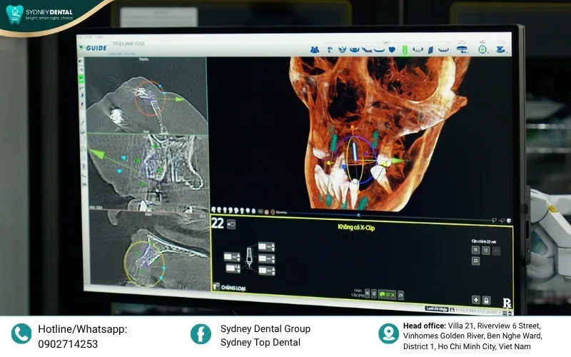
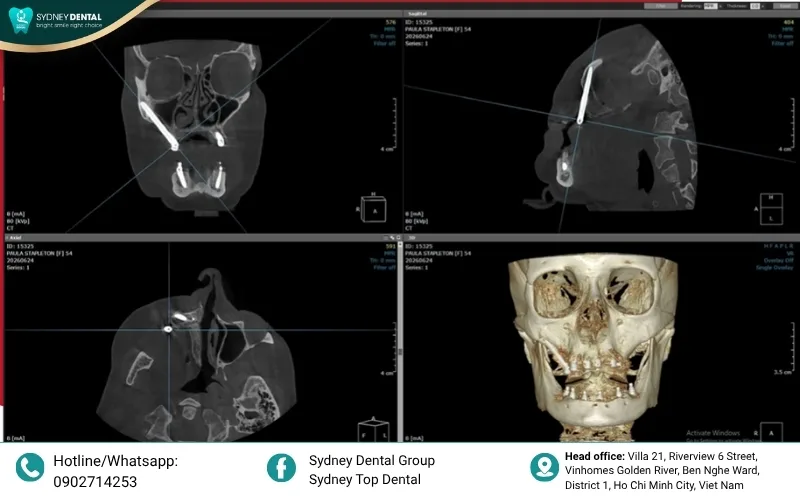
Precision planning using high-resolution CT Cone Beam imaging is the foundation of a safe zygomatic procedure. This technology allows the surgeon to visualize the bone density and anatomical boundaries in three dimensions before the first incision. Accurate mapping of the sinus and nerve pathways is essential to minimize the risk of accidental injury.
Choosing a highly skilled surgeon who utilizes modern techniques like the ZAGA (Zygoma Anatomically Guided Approach) significantly improves patient outcomes. These methods focus on adapting the implant placement to the patient’s unique anatomy rather than using a one-size-fits-all strategy. Using advanced technologies reduces surgical time and enhances the long-term stability of the implants.
Strict adherence to postoperative care instructions, including sinus precautions and oral hygiene protocols, is vital for healing. Patients must avoid smoking and follow a soft-food diet to prevent excessive mechanical stress during the initial integration phase. Regular follow-up appointments allow the clinical team to monitor the site and intervene early if any issues arise.
6. Zygomatic Implants: Minimizing Complications and Optimizing Outcomes by Dr. Hai
For over 20 years, clinical methods for zygomatic implants with immediate loading have evolved to improve safety and effectiveness. Traditional techniques, such as the intrasinus approach or extensive bone grafting, often resulted in specific biological challenges. These included peri-implantitis, maxillary sinusitis, and implant neck exposure due to insufficient soft tissue or poor implant design.
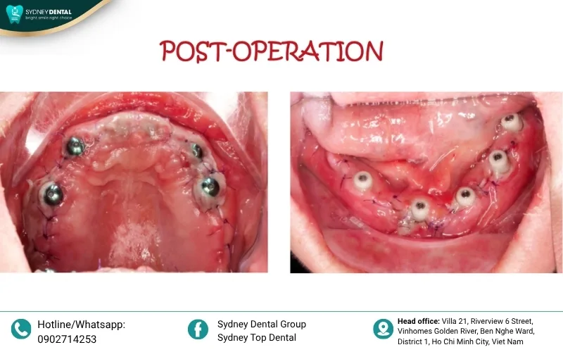
Statistics show that maxillary sinusitis remains a significant concern, affecting over 14% of cases involving immediate loading. To address this, specialized approaches like the Zygoma Anatomically Guided Approach (ZAGA) have been developed by international experts. Dr. Hai has integrated these trends by implementing a minimally invasive technique that prioritizes the protection of the sinus lateral wall.
- Sinus Membrane Protection: Utilizing sinus lift techniques combined with PRF (Platelet-Rich Fibrin) buffers to minimize the risk of membrane perforation.
- Soft Tissue Augmentation: Incorporating connective tissue grafts at the implant neck to increase the thickness of keratinized tissue.
- Reduced Invasiveness: Minimizing trauma to the sinus wall to prevent postoperative inflammatory responses.
By combining these two advanced techniques during the surgical phase, Dr. Hai aims to significantly reduce the incidence of sinusitis. This comprehensive approach ensures that patients can enjoy the benefits of immediate loading with much lower risk. These clinical insights serve as a valuable reference for practitioners seeking to optimize their zygomatic implant results.
Learn more: Zygoma Zaga Centers: A Breakthrough Solution for Patients with Severe Maxillary Atrophy
Zygomatic implants remain a revolutionary “last resort” solution that can transform the lives of patients with severe bone loss. However, achieving success requires a combination of advanced surgical technology, expert skill, and diligent postoperative care. Always remember that for a procedure this complex, choosing the most qualified specialist is the best way to ensure a safe and lasting smile.
Sydney Top Dental
📍 Address: No. 21, Riverview 6 Street, Vinhomes Golden River Villas, Ben Nghe Ward, District 1, Ho Chi Minh City
📞 Hotline: 090 271 42 53
🌐 Facebook: Sydney Top Dental


Our Doctors
Get a Free Initial Consultation